
From Colorless to Blue to Yellow: The Scientific Principles Behind TMB Color Development Reaction
Those who have performed ELISA, Western blot, or biochemical assays are certainly familiar with TMB color development. A transparent, colorless liquid gradually reveals a clear blue after sample addition and incubation. Different shades of blue correspond to the amount of target substance in the sample. Once stop solution is added, it instantly turns uniformly bright yellow. This classic color change sequence from colorless → blue → yellow is the core signal source for quantitative and qualitative detection in countless biological experiments. However, most people only know the operational steps, but have never truly understood the underlying scientific logic.
Why is TMB initially colorless? Why does it definitely turn blue after catalysis? Why does stopping the reaction specifically turn it yellow? Today, we will set aside obscure formulas and break down the complete principles of TMB color development reaction in an easy-to-understand way, revealing the chemical essence behind the experimental phenomena.
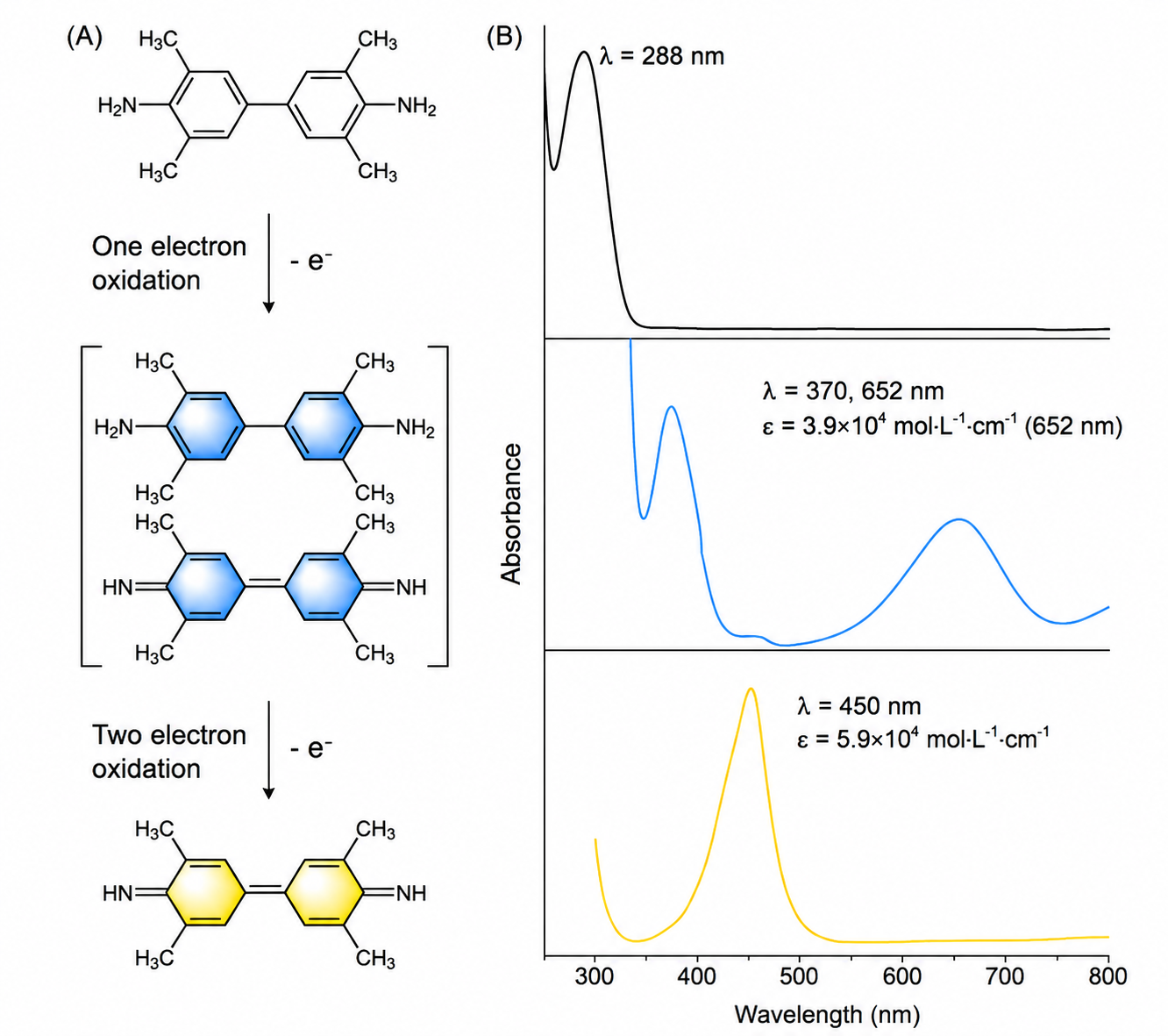
(A) Reaction mechanism of horseradish peroxidase (HRP) catalyzed H₂O₂ oxidation of TMB; (B) Spectral changes of TMB before and after oxidation.
1. Introduction to TMB: The Color-Changing Indicator in Experiments
TMB, short for 3,3',5,5'-tetramethylbenzidine, is the most commonly used highly sensitive chromogenic substrate in biological detection, especially compatible with HRP catalytic systems, and widely used in ELISA kits, colloidal gold, immunohistochemistry, and other experimental scenarios. Its core characteristic is that the oxidation-reduction state determines the color change, and the reaction is mild, highly sensitive, and has low background interference, fully meeting the detection needs of trace biological samples. TMB that has not undergone any reaction is in a reduced state with a stable molecular structure and no chromophore, so the solution appears completely transparent and colorless, which is also the initial form of the TMB chromogenic solution we receive.
2. Colorless to Blue: Signal Generation Through Dynamic Catalysis
After incubation starts, the process of colorless TMB turning blue is essentially an oxidation reaction catalyzed by HRP enzyme, and is also the core stage of experimental signal accumulation. The entire reaction system contains three core elements: substrate TMB, oxidant hydrogen peroxide (H₂O₂), and catalyst HRP enzyme — all three are indispensable.
Under normal conditions, hydrogen peroxide cannot directly oxidize TMB rapidly, while HRP enzyme efficiently activates hydrogen peroxide, causing it to release reactive oxygen species, which then triggers the stepwise oxidation of TMB: First, the reduced colorless TMB molecule loses a single electron to generate a TMB cation radical. This intermediate is extremely unstable and immediately combines with unoxidized colorless TMB molecules to form a dimeric charge-transfer complex, which is the blue soluble product we see in experiments, with maximum absorption at 370nm and 652nm. As incubation time increases, more and more TMB molecules are oxidized, the concentration of charge-transfer complexes continues to rise, and the blue color of the solution becomes deeper and deeper. The depth of blue is positively correlated with HRP activity in the sample and the concentration of target antigen/antibody. Higher sample content leads to more vigorous catalytic reaction, more oxidized TMB, and deeper blue — this is also the core principle enabling ELISA quantitative detection.
3. Blue to Yellow: Structural Stabilization Through Acidic Termination
In experiments, we do not let the blue deepen indefinitely. After incubation, we must add acidic stop solution, and the blue solutions of varying depths will instantly turn uniformly bright yellow. The essence of this color mutation is that the acidic environment completely changes the molecular structure of TMB oxidation products.
In the weakly acidic incubation system, TMB only undergoes single-electron oxidation to generate blue charge-transfer complexes. After adding acid stop solution, the pH of the system drops sharply, promoting all incompletely oxidized TMB intermediates to undergo complete two-electron oxidation, generating stable diimine-type oxidation products. This final steady-state product appears bright yellow in acidic environment, with a characteristic absorption peak at 450nm, which is also the standard wavelength for final readings on laboratory microplate readers. Compared to the unstable blue intermediates of varying depths, the yellow final product has a stable structure, uniform signal, and controllable values, effectively terminating the continuous catalytic reaction of the enzyme, avoiding experimental deviations caused by incubation time errors, and making detection data more accurate and reproducible.
4. Complete Color Change Logic: Understand the Full Process with One Flow Diagram
To facilitate memory, we have organized the complete reaction chain of TMB color development:
Colorless (Initial Reduced TMB) → HRP+H₂O₂ Catalysis, Single-Electron Oxidation → Blue (Charge-Transfer Complex Intermediate) → Strong Acid Termination, Complete Two-Electron Oxidation → Yellow (Stable Diimine Final Product)
The entire process has no complex side reactions, and color changes strictly correspond to molecular oxidation degree and environmental pH. This is also the key reason why TMB has become the mainstream chromogenic substrate — specific reaction, linear signal, and strong controllability.
5. Final Thoughts: Scientific Logic in Tiny Color Changes
From colorless to blue, and then from blue to yellow, the three-color changes of TMB seem simple, but they are actually a precise and rigorous oxidation-reduction regulation system. It is not only the "visual signal" of biological detection but also the perfect combination of chemical structure, enzymatic catalysis, and environmental regulation. Scientific experiments are never mechanical pipetting, incubation, and termination. Every color change and every numerical fluctuation has corresponding scientific principles behind it.
Understanding TMB color development principles not only helps you avoid experimental errors and improve data accuracy but also allows you to truly understand the underlying logic of ELISA and other detection experiments, advancing from "knowing how to do experiments" to "understanding experiments."