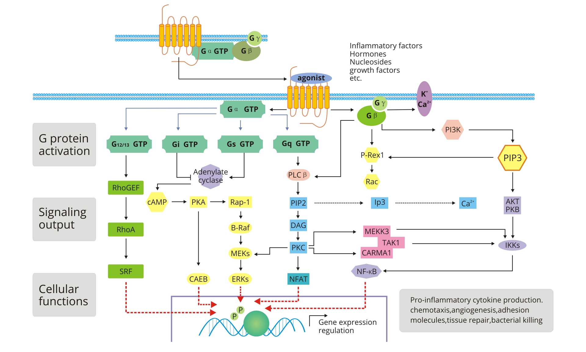
G Protein Coupled Receptor Signaling Pathway
What Are G Protein-Coupled Receptors?
G protein-coupled receptors (GPCRs) are a large family of cell surface receptors that bind and interact with intracellular heterotrimeric GTP-binding proteins to transduce extracellular stimuli into intracellular signals. These external stimuli cover a wide range of signaling molecules, including classic biogenic amines such as adrenaline, norepinephrine, histamine and 5-hydroxytryptamine, as well as peptide hormones including bradykinin, luteinizing hormone and parathyroid hormone. Additionally, olfactory molecules and light signals can also serve as activating ligands for GPCRs. Structurally, GPCRs feature seven transmembrane alpha-helices, linked by short extracellular and intracellular loops, which earns them the alternative name of 7-transmembrane (7-TM) receptors.
What Is the GPCR Signaling Pathway?
GPCRs can be triggered by diverse extracellular ligands, including hormonal factors, growth factors and inflammatory mediators. Upon ligand binding, activated GPCRs function as guanine nucleotide exchange factors (GEFs) for downstream G proteins. They facilitate the conversion of GDP to GTP on G protein subunits, which induces full G protein activation. Activated G proteins dissociate into independent Gα subunits and Gβγ dimers, each of which modulates distinct downstream effector molecules. This sequential signaling cascade amplifies extracellular signals across the cell membrane and triggers a broad spectrum of biological outcomes, such as cell proliferation, survival, differentiation, migration, extracellular matrix remodeling, angiogenesis and tumor progression.
Gα protein subunits are classified into four major subfamilies: Gαs, Gαi, Gαq/11 and Gα12/13. Each subfamily initiates unique downstream signaling cascades and mediates different cellular responses.
Gαs Signaling Branch
The Gαs subunit enhances the catalytic activity of adenylate cyclase (AC), which promotes the conversion of ATP into cyclic adenosine monophosphate (cAMP). Elevated cAMP levels further activate protein kinase A (PKA), triggering downstream signaling cascades including Rap-1/B-Raf/MEKs/ERKs and PKA/CREB pathways to modulate cellular gene transcription.
Gαi Signaling Branch
In contrast, the Gαi subunit inhibits adenylate cyclase activity, reduces intracellular cAMP concentration, and thereby suppresses cAMP-dependent signaling transduction.
Gαq/11 Signaling Branch
The Gαq/11 subunit primarily activates plasma membrane-bound phospholipase C-β (PLC-β). Activated PLC-β hydrolyzes phosphatidylinositol 4,5-bisphosphate (PIP2), a key membrane lipid, to generate two secondary messengers: diacylglycerol (DAG) and inositol trisphosphate (IP3).
IP3 migrates to the endoplasmic reticulum membrane and binds to IP3-sensitive calcium channels, opening these channels and triggering massive calcium ion release from the endoplasmic reticulum into the cytoplasm. The accumulated cytoplasmic calcium ions bind and recruit inactive protein kinase C (PKC) to the cell membrane, where PKC interacts with DAG to achieve full activation. Activated PKC phosphorylates multiple downstream signaling substrates to regulate diverse cellular behaviors, including cellular secretion, muscle contraction, cell proliferation and cell differentiation.
IP3 migrates to the endoplasmic reticulum membrane and binds to IP3-sensitive calcium channels, opening these channels and triggering massive calcium ion release from the endoplasmic reticulum into the cytoplasm. The accumulated cytoplasmic calcium ions bind and recruit inactive protein kinase C (PKC) to the cell membrane, where PKC interacts with DAG to achieve full activation. Activated PKC phosphorylates multiple downstream signaling substrates to regulate diverse cellular behaviors, including cellular secretion, muscle contraction, cell proliferation and cell differentiation.
Gα12/13 & Gβγ Regulatory Function
The Gα12/13 subunits regulate the activity of the small GTPase RhoA by interacting with Rho guanine nucleotide exchange factors (RhoGEFs). Both Gα12 and Gα13 can activate PDZ-RhoGEF, while Gα13 directly binds and stimulates p115RhoGEF, ultimately promoting RhoA-dependent signaling activation.
Separated Gβγ dimers also participate in multiple signaling regulations. They modulate the opening and closing status of cellular potassium and calcium ion channels. Moreover, Gβγ subunits recruit multiple signaling proteins to the plasma membrane, such as PI3K, P-Rex-1 and β-adrenergic receptor kinase. Most downstream effector pathways are synergistically regulated by both Gα subunits and Gβγ dimers.
Separated Gβγ dimers also participate in multiple signaling regulations. They modulate the opening and closing status of cellular potassium and calcium ion channels. Moreover, Gβγ subunits recruit multiple signaling proteins to the plasma membrane, such as PI3K, P-Rex-1 and β-adrenergic receptor kinase. Most downstream effector pathways are synergistically regulated by both Gα subunits and Gβγ dimers.
Biological Functions of the GPCR Signaling Pathway
GPCR signaling networks participate in a wide array of fundamental physiological processes. These include visual and olfactory signal perception, behavioral and emotional regulation, immune response modulation, autonomic nervous system homeostasis, cell density sensing and overall physiological balance maintenance.
Association Between GPCR Signaling and Human Diseases
Given the extensive regulatory roles of GPCRs in nearly all physiological activities, abnormal GPCR signaling transduction is closely associated with human diseases and cellular functional disorders.
Dysregulated GPCR signaling is commonly observed in multiple pathological conditions, including cardiovascular disorders, hereditary diseases, malignant tumors and infectious diseases.
For instance, cholera toxin impairs the GTPase activity of G protein α subunits, leading to persistent adenylate cyclase activation. Continuously activated AC generates excessive cAMP, which opens plasma membrane ion channels, causes massive leakage of intracellular ions and water, and ultimately leads to severe cellular dehydration.
Dysregulated GPCR signaling is commonly observed in multiple pathological conditions, including cardiovascular disorders, hereditary diseases, malignant tumors and infectious diseases.
For instance, cholera toxin impairs the GTPase activity of G protein α subunits, leading to persistent adenylate cyclase activation. Continuously activated AC generates excessive cAMP, which opens plasma membrane ion channels, causes massive leakage of intracellular ions and water, and ultimately leads to severe cellular dehydration.
Genetic mutations in GPCR genes can result in a variety of congenital and acquired diseases, such as retinitis pigmentosa, hyperthyroidism, renal diabetes insipidus and reproductive system disorders. Accumulating research evidence demonstrates that aberrant GPCR signaling is tightly correlated with tumor initiation and progression. Multiple GPCR subtypes are abnormally overexpressed in various malignancies. For example, LPA receptors show high expression levels in ovarian, breast, colon and prostate cancers, while endothelin receptors are significantly upregulated in colon cancer, prostate cancer and melanoma.