
The cell division cycle (shortened as cell cycle) refers to the full recurring process of eukaryotic proliferative cells from the completion of one cell division to the start of the next. During this whole process, cellular chromosomal DNA undergoes complete accurate duplication, followed by even partitioning of replicated genetic material into two independent daughter cells with identical genetic compositions.
As a foundational biological process, the cell cycle supports embryonic development starting from a single fertilized zygote into a mature complete organism. It also maintains steady physiological tissue renewal, supporting continuous regeneration of epidermal cells, hair follicles, blood cells and multiple internal organ tissues.
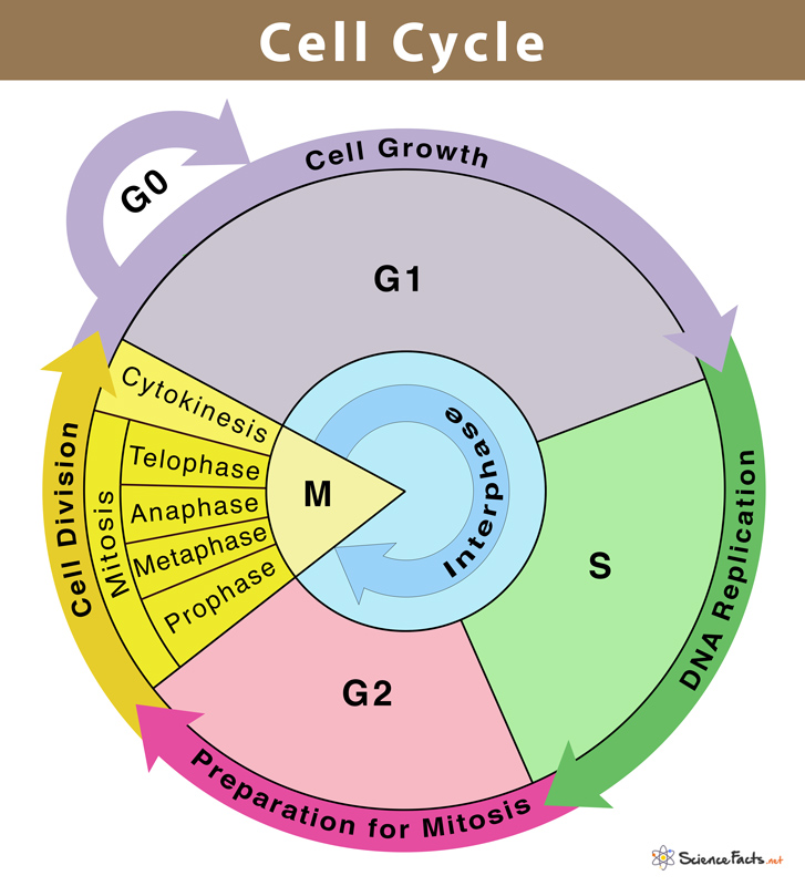
All eukaryotic cells with nuclei follow a two-part cell cycle framework: interphase and mitotic (M) stage. Interphase takes up roughly 91% to 95% of the entire cycle duration, serving as the preparation interval ahead of actual cell division. It can be further split into three sequential sub-stages: G1 phase, S phase and G2 phase.
2.1 Interphase
G1 Phase (Gap 1)
This initial growth stage immediately follows the previous mitotic division. Cells sustain vigorous metabolic activity without genomic DNA replication. Large-scale synthesis of mRNA and functional proteins takes place to lay material foundations for subsequent DNA duplication. Cells simultaneously sense extracellular growth cues and internal nutrient reserves to decide whether to enter active proliferation or shift to the dormant G0 resting state.
S Phase (Synthesis Stage)
The core stage for genomic DNA replication. The double-stranded DNA of every chromosome is fully duplicated to form two identical sister chromatids connected at the centromere. If the initial ploidy is 2N, the total DNA content rises to 4N after replication, while the chromosome quantity stays unchanged. Apart from DNA strands, histone proteins required for new chromatin assembly are also produced within this period.
G2 Phase (Gap 2)
The final preparation stage prior to mitosis. Centriole pairs finish full duplication, and massive synthesis of tubulin and mitosis-associated regulatory factors occurs. Cells carry rigorous genomic damage monitoring; any unrepaired DNA lesions will stall the cell cycle at the G2 checkpoint and prevent faulty mitosis from proceeding.
2.2 M Phase (Mitosis & Cytokinesis)
The M stage consists of nuclear mitosis and subsequent cytoplasmic separation (cytokinesis). Mitosis is divided into four ordered sub-stages: prophase, metaphase, anaphase and telophase.
- Prophase: Loose chromatin condenses into compact, visible chromosomes; the nuclear membrane and nucleolus break down. Spindle microtubules assemble from duplicated centrioles and extend toward chromosomes.
- Metaphase: Spindle fibers firmly attach to the kinetochore on each chromosome’s centromere, pulling all chromosomes to neatly align on the cell’s central equatorial plane.
- Anaphase: Centromeres fully split apart, separating paired sister chromatids. Spindle microtubules pull individual chromatids toward two opposite cell poles, doubling the cell’s total chromosome number temporarily.
- Telophase: New nuclear envelopes reform around two groups of separated chromosomes, and nucleoli regenerate. Chromosomes gradually decondense back into loose chromatin, while the mitotic spindle apparatus disassembles.
After telophase finishes, cytokinesis initiates to separate cytoplasm and organelles into two separate daughter cells. Cytokinesis mechanisms differ greatly between animal and plant cells:
- Animal cells: An actin-myosin contractile ring forms at the cell equator and constricts inward to form a cleavage furrow, splitting the cell into two identical progeny cells.
- Plant cells: Due to rigid cellulose cell walls, no contractile ring forms. Instead, Golgi-derived vesicles fuse at the cell midline to form a cell plate, which expands outward and matures into new cell walls to divide the parent cell.
Precise, ordered molecular signaling cascades tightly control cell cycle progression, including DNA damage sensing-repair pathways and inhibitory mechanisms for uncontrolled cell proliferation. All phase transitions depend on the coordinated activation or degradation of core regulatory protein complexes.
3.1 Core Regulators: Cyclins & Cyclin-Dependent Kinases (CDKs)
Cyclins are phase-specific regulatory proteins with fluctuating expression levels throughout the whole cycle. They bind and activate cyclin-dependent kinases (CDKs) to form functional heterodimer enzymes. Activated cyclin-CDK complexes phosphorylate downstream target proteins to drive cell cycle transition at G1/S, G2/M and mitotic checkpoints:
- Cyclin D/CDK4/6: Regulates early G1 progression and G1-S checkpoint passage
- Cyclin E/CDK2: Controls late G1 advancement and triggers DNA replication entry
- Cyclin A/CDK2: Maintains S-phase DNA synthesis and early G2 progression
- Cyclin B/CDK1 (MPF): Initiates G2/M transition and coordinates the whole mitotic process
3.2 CDK Inhibitors (CKIs)
Two major protein families suppress CDK activity under stress or DNA damage signals to arrest cell cycle progression:
- INK4 family (p16, p15, p18): Specifically bind CDK4/6 to sequester Cyclin D complexes and lock cells in G1 phase
- CIP/KIP family (p21, p27): Broad-spectrum suppressors that block all cyclin-CDK complexes to pause the cycle at multiple checkpoints
3.3 Cell Cycle Checkpoint Surveillance Systems
Three vital checkpoints guarantee genomic stability:
- G1 Checkpoint: Examines cell volume, nutrient availability, growth signals and DNA lesions; defective cells enter G0 state or activate repair pathways
- G2 Checkpoint: Scans for incomplete DNA replication or post-replication damage before mitosis starts
- Spindle Assembly Checkpoint (Metaphase Checkpoint): Confirms all chromosomes are correctly anchored to spindle fibers before anaphase launches
3.4 Ubiquitin-Dependent Protein Degradation Pathway
The Anaphase-Promoting Complex/Cyclosome (APC/C) functions as a key E3 ubiquitin ligase. It labels cyclins and securin proteins with ubiquitin tags for proteasome breakdown, enabling sister chromatid separation in anaphase and exit from mitosis.
Disordered cell cycle control leads to unrestrained cell proliferation, which acts as the core driving factor of tumor formation. Mutations that break cyclin-CDK balance, inactivate CKIs or damage checkpoint monitoring allow cells carrying mutated genomes to bypass cycle arrest and replicate continuously.
- Oncogenic variations: Overexpression of Cyclin D1/2/3, CDK4/6 gene amplification, loss-of-function mutations of p16/p21
- Tumor suppressor defects: Mutated p53 (the master DNA damage sensor) abolishes G1 and G2 checkpoint arrest upon genomic injury
Apart from malignant tumors, irregular cell cycle progression also contributes to degenerative diseases, cellular senescence and developmental malformations caused by disordered embryonic cell proliferation timing.