
Rabies virus (RABV) belongs to the Lyssavirus genus under the Rhabdoviridae family, a neurotropic pathogen that triggers fatal encephalitis across all warm-blooded animal species. Globally, domestic dogs act as the primary natural reservoir and transmission vector of RABV. Human infection mainly occurs when virus-laden saliva from infected animals enters the body via bites or scratches. Timely administration of rabies vaccine combined with human rabies immunoglobulin after exposure is critical to block disease onset, yet there are no targeted antiviral medications capable of directly neutralizing rabies virus particles.
RABV presents a characteristic bullet-shaped enveloped virion, with an average length of roughly 180 nm and a diameter of approximately 75 nm. The outermost layer consists of a lipid bilayer envelope studded with spike-shaped glycoprotein (G) molecules. Beneath the envelope lies the matrix protein (M) layer; internally, nucleoprotein (N) tightly wraps viral genomic RNA to form the core nucleocapsid. This helically symmetric ribonucleoprotein complex (RNP) is further assembled with phosphoprotein (P) and large polymerase protein (L).
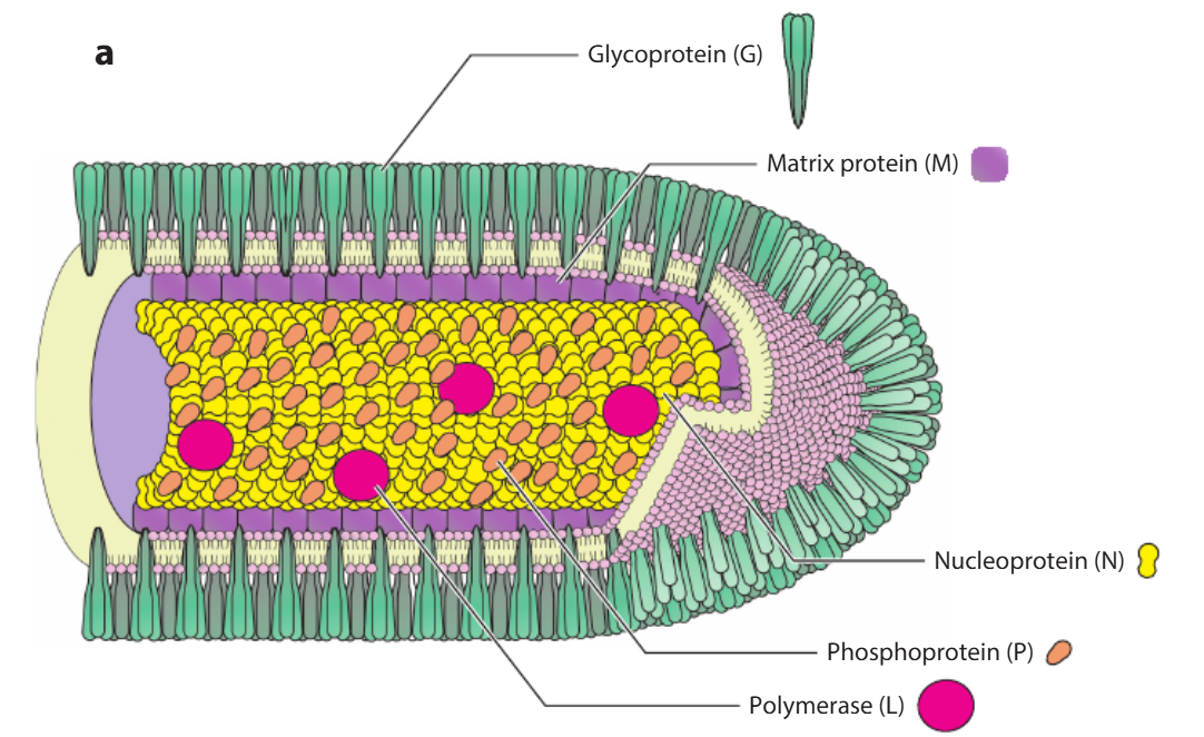
Figure 1. Schematic diagram of RABV structure
RABV carries a non-segmented single-stranded negative-sense RNA genome spanning about 11.9 kb. This viral genome encodes five distinct structural proteins: nucleoprotein (N), phosphoprotein (P), matrix protein (M), glycoprotein (G), and RNA-dependent RNA polymerase (L).
N protein encapsidates viral RNA to form the ribonucleoprotein (RNP) complex. Together with L and P proteins, RNP condenses into a helical nucleocapsid (NC). P protein functions as an essential non-catalytic cofactor for the L polymerase. M protein participates in viral budding, host cell apoptosis induction, and intracellular membrane rearrangement. Trimeric G protein interacts with M protein through its cytoplasmic tail; it is the sole surface-exposed protein on rhabdovirus envelopes and serves as the exclusive ligand binding to host cell receptors, mediating viral attachment and cellular entry.
Table: Functional Introduction of RABV Structural Proteins
| RABV Structural Proteins | Core Biological Function |
|---|---|
| Nucleoprotein (N) | Combines with viral RNA to construct nucleocapsid |
| Phosphoprotein (P) | Regulates viral RNA synthesis and genome replication |
| Matrix Protein (M) | Governs viral particle assembly and membrane budding |
| Glycoprotein (G) | Anchored on viral envelope, mediates host cell adhesion and invasion |
| RNA-dependent RNA polymerase (L) | Indispensable for viral genome replication and gene transcription |
RABV infection starts with virion attachment to host cell surfaces, followed by internalization via endosomal trafficking pathways. After entering host cytoplasm, the virus synthesizes all required structural proteins and genomic RNA for progeny virion assembly, which are eventually released outward through membrane budding.
2.1 Viral Entry Process
RABV invades target cells via clathrin-dependent endocytosis. Surface G protein mediates binding to host cell membranes, then triggers membrane fusion to deliver viral contents into the cytoplasm. After internalization, G protein drives pH-sensitive fusion with endosomal membranes, leading to viral uncoating and release of helical nucleocapsid (NC) from the RNP complex.
2.2 Viral Genome Replication
The five gene segments (N, P, M, G, L) on nucleocapsid genomic RNA are transcribed into five independent monocistronic mRNAs by virion-bound RNA-dependent RNA polymerase. Each mRNA undergoes translation to generate corresponding N, P, M, G and L proteins, which supply raw materials for new virion assembly. After sufficient viral protein accumulation, genome replication proceeds: full-length positive-sense RNA is generated as a template for producing progeny negative-sense viral RNA.
2.3 Virion Assembly & Extracellular Release
Newly replicated genomic RNA is encapsidated by N-P complexes, which recruit L protein to form nascent RNP cores. M protein binds to these RNP cores to compact them into stable scaffold structures. These scaffolds interact with trimeric G proteins embedded in the host plasma membrane to assemble complete viral particles. Mature virions bud off from infected cell membranes into extracellular interstitial fluid.
Rabies disease is triggered by RABV infection, and the pathogen spreads mainly through saliva secreted by infected animals. Bites from infected animals constitute the primary transmission channel to humans or other animals. All mammal species are theoretically capable of transmitting rabies virus; high-risk species that frequently infect humans include dogs, cats, cattle, ferrets, goats, horses, bats, beavers, coyotes, foxes, monkeys, raccoons, skunks and groundhogs.
In rare circumstances, infection may occur when virus-containing saliva contacts broken skin or mucosal tissues such as oral and ocular surfaces, for instance when an infected animal lacerates or licks open wounds on human skin.
A large body of research confirms that rabies pathogenic progression, covering incubation and clinical onset phases without detectable viremia, can be divided into three distinct stages.
4.1 Local Tissue Proliferation Stage
After invading through bite wounds, RABV accumulates and replicates within nerve fibers of striated muscle spindle receptors at the injury site, then migrates into adjacent peripheral nerve tissue. The interval from local colonization to peripheral nerve invasion usually lasts less than three days, while some research indicates the virus may persist at the wound site for up to two weeks or longer.
4.2 Central Nervous System Invasion Stage
RABV travels centripetally along peripheral nerve axoplasm at an approximate speed of 3 mm per hour. Upon reaching dorsal root ganglia, the virus undergoes massive amplification, then spreads into the spinal cord and the entire central nervous system, preferentially infecting neurons distributed in the cerebrum and cerebellum.
4.3 Systemic Organ Diffusion Stage
After replicating within the central nervous system, the virus spreads centrifugally via peripheral nerves to invade multiple tissues and organs, with prominent tropism for salivary nuclei, glossopharyngeal nuclei and hypoglossal nuclei.
Recent rabies-related studies have achieved breakthroughs across multiple research dimensions. Importation risks have drawn widespread attention, with documented cases of canine RABV variants carried by dogs imported into the United States from high-prevalence regions including India, Iraq and Egypt. Studies on RABV molecular biology have identified key host factors that regulate viral replication efficiency. Diagnostic technology innovations include a fast, sensitive reverse transcription recombinase polymerase amplification assay suitable for resource-limited regions to detect rabies virus.
Novel recombinant vaccines based on chimpanzee adenovirus vector (ChAd68-Gp) have induced robust immune responses and provided effective protection against lethal viral challenge in beagle models. Research advances have also been reported for mRNA vaccines encoding the primary RABV antigen G protein. Multiple clinical trials have evaluated optimized vaccination schedules and streamlined visit protocols, verifying sustained protective neutralizing antibody titers in vaccinated populations.
Field surveillance on common vampire bats in Mexico recorded multiple mortality outbreaks, revealing extended incubation cycles and dynamic changes in post-infection neutralizing antibody levels. Additionally, a Knowledge, Attitude and Practice (KAP) survey conducted in Uganda supplied valuable epidemiological data on local rabies prevalence and completed molecular typing of circulating RABV strains. Collectively, these research outputs deepen our understanding of rabies covering import risk prevention, diagnostic technology, vaccine development and population epidemiology.