
Inflammasome Signaling Pathway
What Is An Inflammasome?
Inflammasomes are core cytosolic multiprotein oligomeric complexes that serve as pivotal sensory and effector machineries of the mammalian innate immune system. They are predominantly assembled in myeloid cells including macrophages, dendritic cells, and microglia, and can also be induced in epithelial cells and neurons under stress conditions. Structurally, a canonical inflammasome complex consists of three core functional components: an upstream pattern recognition receptor (PRR) sensor protein belonging to the NOD-like receptor (NLR) family or the AIM2-like receptor (ALR) family, the central adaptor protein ASC (apoptosis-associated speck-like protein containing a C-terminal CARD domain), and the downstream effector protease pro-caspase-1. As a crucial intracellular immune surveillance system, inflammasomes are specifically responsible for detecting endogenous danger signals and exogenous pathogenic stimuli, bridging the gap between pathogen recognition, cellular stress sensing, and downstream inflammatory immune responses. Unlike membrane-bound pattern recognition receptors, inflammasomes function in the cytoplasm, enabling the host to monitor intracellular infections, tissue damage, and metabolic abnormalities that cannot be identified by cell surface receptors. Multiple distinct inflammasome subtypes have been identified in humans and mice, among which NLRP1, NLRP3, NLRC4, and AIM2 inflammasomes are the most well-characterized, each exhibiting unique ligand specificity and tissue distribution to cope with diverse immune challenges.
The Function of Inflammasome Signaling
Inflammasome signaling acts as a double-edged sword in regulating host immune homeostasis, physiological repair, and pathological inflammation, participating in the entire process of innate immune defense and tissue remodeling. Physiologically, inflammatory responses triggered by inflammasome activation are essential components of the body’s self-healing and defensive mechanisms. Upon exposure to microbial invasion, cellular oxidative stress, tissue trauma, metabolic dysfunction, or toxic substance stimulation, inflammasome complexes rapidly assemble and initiate downstream signaling cascades to eliminate harmful stimuli and repair damaged tissues. In peripheral immune cells, inflammasome activation mediates the programmed inflammatory cell death termed pyroptosis, which specifically eliminates pathogen-infected, damaged, or functionally abnormal immune cells, thereby preventing the persistent survival of hazardous cells and limiting the spread of intracellular pathogens. Beyond immune defense, basal inflammasome activity maintains normal tissue metabolism and immune surveillance, regulating cell proliferation, autophagy, and tissue microenvironment stability.
Pathologically, abnormal or excessive activation of inflammasome signaling is closely associated with multiple human diseases. Sustained inflammasome hyperactivation leads to excessive secretion of pro-inflammatory cytokines and uncontrolled pyroptosis, triggering persistent inflammatory cascades, tissue inflammatory injury, and immune homeostasis imbalance. A large number of studies have confirmed that aberrant inflammasome activity is involved in the occurrence and development of infectious diseases, chronic inflammatory diseases, autoimmune disorders, metabolic diseases, and neurodegenerative diseases. In particular, age-dependent upregulation of inflammasome activity drives chronic neuroinflammation and neuronal senescence, further inducing neuropsychiatric disorders such as depression, anxiety, and cognitive decline. In the central nervous system, although neurons can secrete a small amount of IL-1 family inflammatory cytokines, activated microglia and astrocytes are the dominant cellular sources of these cytokines, and their inflammasome hyperactivation is the core driver of neuroinflammatory damage.
The Mechanism of Inflammasome Signaling Pathway
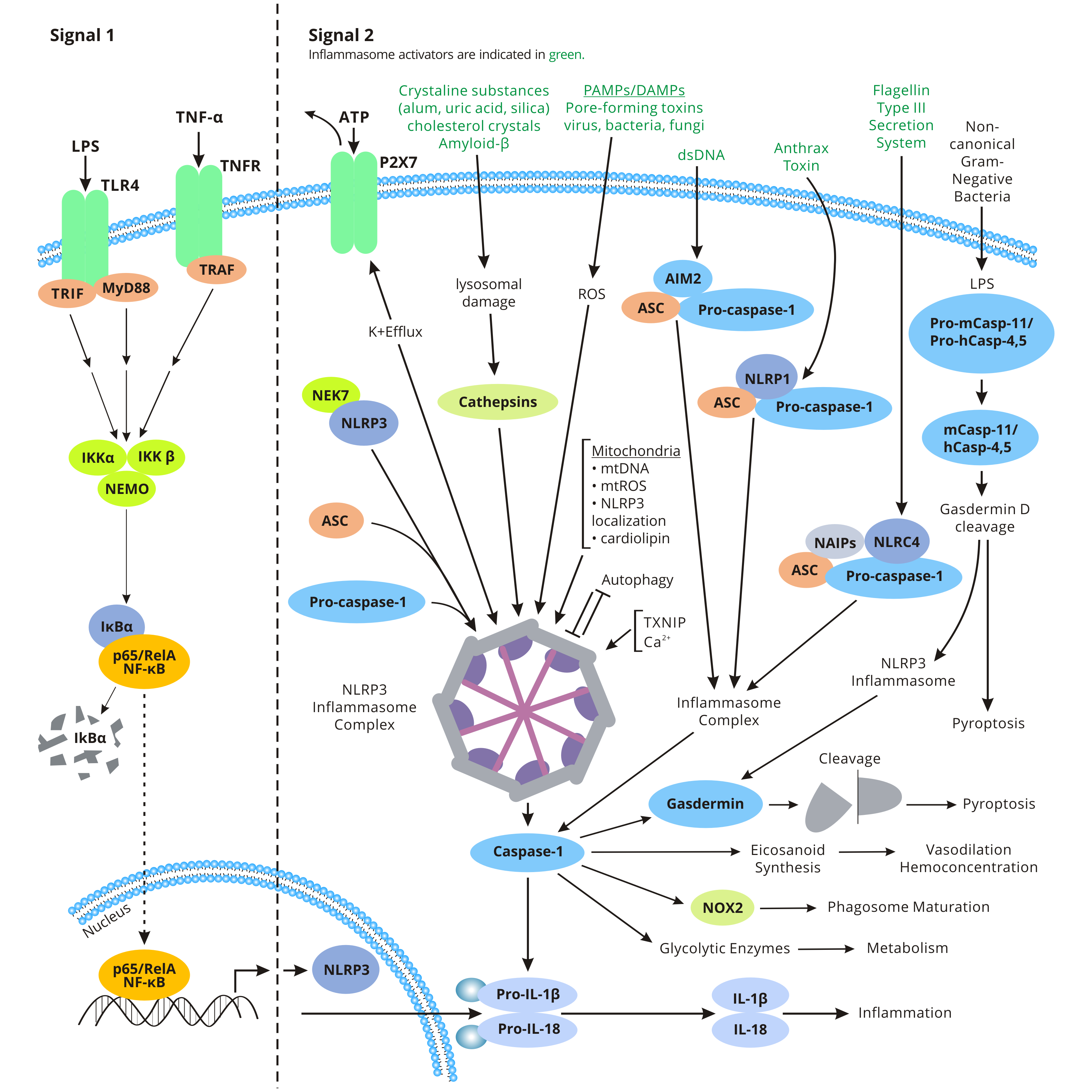
Activated by a variety of PAMPs (pathogen-associated molecular patterns) and DAMPs (damage-associated molecular patterns), the formation of inflammasomes is initiated by the NLR proteins such as NLRP1 , NLRP2 , NLRP3, NLRP6, NLRP7, NLRC4, and the AIM2 (absent in melanoma-2).
A number of inflammasomes have been recognized based on molecular structure such as NLRP1, NLRP2, NLRP3, NLRC4, and AIM2. The NLRP3 inflammasome, formed by the NLR, the adapter molecule ASC (poptosis-associated speck-like protein containing CARD), and the effector molecule pro-caspase-1, is the most studied and the best characterized. The N-terminal of NLRP3 has a PYD domain. After activation, it undergoes oligomerization through the NOD domain and recruits ASC through the PYD-PYD interaction with the ASC molecule. NLRP3 also a C-terminal CARD region, which can combine with the CARD region of caspase-1 (NLRC4 can recruit caspase-1 directly through its N-terminal CARD region). Through the interaction between these proteins, NLR-PYD-caspase-1, a large molecular weight protein complex termed inflammasome is formed.
Inflammasome assembly leads to the autocleavage of caspase-1. Active caspase-1 triggers the processing of pro-IL-1β and pro-IL-18 into their mature and bioactive forms: IL-1β and IL-18. The maturation and secretion of the proinflammatory cytokines IL-1β and IL-18 induce several biological effects related to inflammation, infection, and autoimmune processes. Activation of inflammasomes can also trigger pyroptosis. This effect usually ruptures the cell membrane perforation of infected cells within a short time and unleashes the cytoplasm containing inflammatory factors such as IL-1B and IL-18 into the interstitial space, activating the corresponding receptors of neighboring cells and causing more large-scale immune response.
Studies have been shown that inflammasome activity increases with age, indicating that inflammasomes are associated with neuroinflammation during neuronal aging. Neuronal aging will result in psychiatric disorders like depression. While neurons also secrete IL-1 family cytokines, activated microglia and astrocytes are the main sources of these cytokines in the brain.
On the other hand, a different set of NLRs, including NLRP1, NLRP3, and NLRC4, induce caspase-1 activation through the assembly of multiprotein complexes called inflammasomes. The activated of caspase-1 regulates maturation of the pro-inflammatory cytokines IL-1β, IL-18 and drives pyroptosis. IL-1β release mediates the activation of the IL-1 receptor signaling pathway and myeloid differentiation factor (MyD88)-dependent nuclear factor kappa B, resulting in the transcription of cytokines such as IL-8, S100, and macrophage inflammatory protein 2 (MIP2), generating cascade amplification effect.
| Targets | ELISA Kits |
|---|---|
| AIM2 | AIM2 ELISA Kit |
| IL18 | IL18 ELISA Kit |
| IL1B | IL1B ELISA Kit |
| LCAT | LCAT ELISA Kit |
| NFKBIA | NFKBIA ELISA Kit |
| Caspase 1 | Caspase 1 ELISA Kit |
| NLRC4 | NLRC4 ELISA Kit |
| TNF-α | TNF-α ELISA Kit |
| LPS | LPS ELISA Kit |
| TXNIP | TXNIP ELISA Kit |