
T lymphocytes serve as core functional components of the immune system, defending the human body against invasive pathogens and malignant tumor cells. Deciphering the sophisticated regulatory network governing T cell differentiation is critical to clarifying the developmental trajectory and biological functions of these immune effector cells. Starting from hematopoietic stem cell generation in bone marrow, followed by thymic maturation and antigen-triggered activation within peripheral tissues, every developmental stage of T cells features distinct shifts in surface biomarker expression and functional potency.
Within this review, we systematically outline key developmental phases of T cell differentiation, elaborate on upstream regulatory cytokines, transcriptional regulators, metabolic cascades and intracellular signaling axes that govern T cell fate commitment. We also summarize canonical surface biomarkers used to distinguish T cell subpopulations, alongside the clinical relevance of aberrant T cell differentiation in immune-related disorders.
T cell differentiation refers to a tightly controlled developmental cascade: bone marrow-residing hematopoietic stem cells (HSCs) differentiate into common lymphoid progenitors (CLPs), which subsequently migrate to the thymus to complete maturation and further branch into functionally specialized T cell subgroups with distinct immune roles.
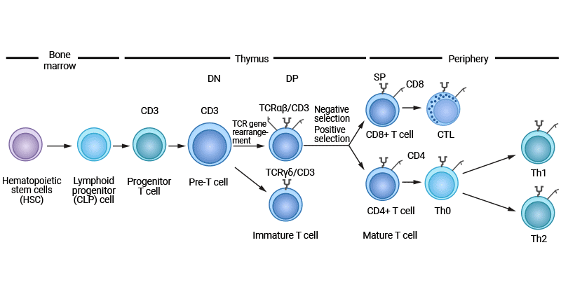
Proper T cell differentiation lays the foundation for robust host immune defense, sustains central and peripheral immune tolerance, and participates in anti-tumor immune surveillance.
The thymus acts as the primary organ for T cell lineage specification and maturation. Bone marrow-derived lymphoid progenitors undergo multi-step developmental transitions inside the thymus to generate antigen-competent mature T lymphocytes.
All T cell lineages originate from multipotent hematopoietic stem cells located in bone marrow. These stem cells first commit to the lymphoid lineage to form CLPs, which circulate via the bloodstream and home to thymic tissue to initiate T cell differentiation programs.
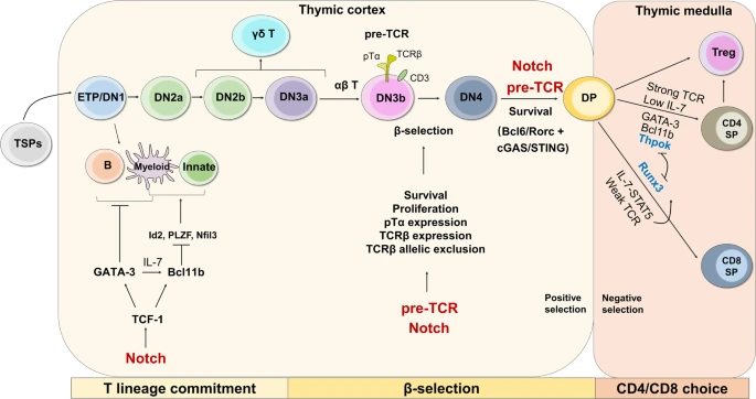
After migrating to the thymic cortex, bone marrow-derived lymphoid progenitors transform into pro-T cells, marking irreversible T lineage commitment mainly initiated by Notch receptor signaling.
DN thymocytes lack surface CD4 and CD8 co-receptors. During this window, thymocytes rearrange the TCR β-chain gene locus. Cells that successfully assemble functional pre-TCR complexes (composed of rearranged TCRβ, pTα and CD3 subunits) undergo β-selection and differentiate into αβ-T cell precursors. Cells failing productive TCRβ rearrangement preferentially develop into γδ T cells.
Thymocytes passing β-selection upregulate both CD4 and CD8 surface markers, entering the DP developmental stage. Both sustained Notch signaling and functional pre-TCR signaling facilitate the DN-to-DP developmental transition.
DP thymocytes undergo positive selection within the thymic cortex, a quality-control process screening TCRs for effective recognition of self-MHC-peptide complexes presented by thymic epithelial cells. Only thymocytes with moderate MHC-binding affinity survive and proceed to subsequent maturation steps.
Post positive selection, DP thymocytes carrying MHC I-restricted TCRs differentiate into CD8+ SP cells, while those with MHC II-restricted TCRs become CD4+ SP cells; this fate bifurcation event is termed CD4/CD8 lineage choice and primarily takes place within the thymic medulla.
SP thymocytes with excessively high TCR affinity for self-antigens undergo apoptotic elimination (negative selection); a minor fraction of high-affinity self-reactive SP cells differentiate into regulatory T cells (Tregs). Surviving low self-reactivity SP cells become fully mature naive T cells and exit the thymus to populate peripheral immune organs.
Mature CD4+ helper T cells and CD8+ cytotoxic T cells traverse the corticomedullary junction to exit the thymus, migrating to secondary lymphoid organs including lymph nodes and the spleen. In peripheral tissues, naive T cells remain quiescent until encountering cognate antigen stimulation.
T cell activation relies on dual signals delivered by antigen-presenting cells (APCs). APCs process exogenous or endogenous antigens and load antigenic peptides onto MHC molecules. Surface TCR complexes on T cells bind to MHC-peptide conjugates to transmit the primary antigen-recognition signal. CD4+ T cells exclusively interact with MHC class II complexes, whereas CD8+ T cells recognize peptides presented by MHC class I molecules.
Antigen-TCR ligation alone cannot achieve full T cell activation. Costimulatory signals provide secondary indispensable stimuli: T cell surface CD28 binds APC-expressed B7 family ligands to amplify intracellular activation cascades. During APC-T cell crosstalk, antigen-presenting cells secrete multiple cytokines such as IL-2 to further boost T cell proliferation and functional differentiation.
Upon successful dual-signal activation, T cells undergo rapid clonal proliferation, generating large pools of identical TCR-expressing clones specific for the triggering antigen. Microenvironmental cytokine signals dictate the terminal effector subtype each activated T cell will differentiate into.
CD8+ T cells polarize into cytotoxic T lymphocytes (CTLs) specialized in lysing virally infected and neoplastic target cells. CD4+ helper T cells branch into multiple functionally distinct subgroups including Th1, Th2 and Th17 subsets. Th1 cells dominate cell-mediated immunity, activating tissue-resident macrophages and enhancing CTL cytotoxicity. Th2 cells orchestrate humoral immune responses by promoting B cell antibody secretion. Th17 cells are central mediators of inflammatory and autoimmune pathologies. Tregs suppress aberrant immune reactivity to preserve self-tolerance and prevent autoimmunity.
Following resolution of immune responses, a portion of effector T cells convert into long-lived memory T cells, enabling accelerated secondary immune responses upon re-exposure to identical antigens.
T cell lineage specification represents a multi-layered regulatory process modulated by diverse inputs: antigen concentration and molecular format, secreted cytokine gradients, lineage-defining transcription factors and local tissue immune microenvironments.
Distinct antigen varieties exert divergent impacts on T cell priming and polarization. Protein antigens serve as robust T cell activators, while non-protein antigens often require carrier molecules to initiate adaptive immune responses. Low antigen dosages typically induce peripheral T cell tolerance, whereas high antigen concentrations robustly trigger T cell activation and downstream differentiation.
Cytokines act as master switches determining CD4+ helper T cell subset polarization. Distinct cytokine cocktails lock T cells into specific effector lineages:
IL-2, IL-12 and IFN-γ collectively drive Th1 polarization to reinforce cellular immunity. IL-4 skews differentiation toward Th2 cells, critical for anti-parasite defense and allergic inflammatory responses. Combined TGF-β and IL-6 signaling promotes Th17 cell development, a population strongly implicated in autoimmune tissue damage.
IL-10 supports Tr1 regulatory cell formation while simultaneously suppressing Th1 and Th2 effector activity to maintain immune equilibrium and limit excessive inflammatory reactions. IL-27 participates in Treg functional maturation and also augments Th1-type immune responses.
Unique transcription factor repertoires govern the expression of subset-specific effector genes in T cell populations. Foxp3 functions as the master transcription factor controlling Treg differentiation and suppressive capacity; fluctuations in Foxp3 expression directly alter Treg development and immune-regulatory functionality. RORγt serves as the core transcriptional driver of Th17 lineage commitment.
TCF1 exerts essential regulatory functions starting at the early thymic progenitor (ETP) stage and is mandatory for initiating T lineage transcriptional programs. Activated downstream of Notch signaling, TCF1 upregulates intermediate transcription factors such as GATA-3 to sustain thymic T cell maturation, regulating all developmental transitions from DN thymocytes to peripheral mature T cells.
GATA-3 directs early T cell development through the DN2 phase by activating T lineage-specific gene networks. Competitive interplay between GATA-3 and T-bet governs CD4+ helper cell polarization: GATA-3 expression favors Th2 formation, while T-bet drives Th1 differentiation.
HES1, a downstream transcriptional effector of Notch signaling, acts during early intrathymic development to block premature differentiation and preserve the undifferentiated state of T cell progenitors.
E protein family transcription factors (E2A, E2-2) in pro-T cells tune stage-specific Notch expression and core T lineage genes, mediate Notch-dependent GATA-3 upregulation, and repress innate lymphoid cell (ILC) developmental programs within the thymus to solidify T cell identity.
Varied tissue niches and pathological conditions (chronic inflammation, solid tumor lesions) reshape T cell developmental trajectories and functional output. Tumor microenvironments secrete inhibitory cytokines and activate immunosuppressive signaling cascades to blunt T cell effector function, facilitating tumor immune evasion.
T cell differentiation is tightly coupled to dynamic metabolic reprogramming, with interconnected metabolic and signaling pathways dictating cellular fate at every developmental stage. Each T cell subset displays a characteristic metabolic signature matched to its energy and biosynthetic demands.
Notch and IL-7 signaling represent the primary metabolic regulators during intrathymic DN, DP and SP maturation. Notch signaling upregulates aerobic glycolysis to supply sufficient biosynthetic precursors and energy for thymocyte proliferation and differentiation. IL-7 signals through the JAK-STAT axis to boost glycolytic flux and fatty acid oxidation, supporting thymocyte growth and survival.
SP thymocytes dynamically toggle between glycolysis and fatty acid oxidation to balance energy supply and nutrient availability, gaining heightened sensitivity to extracellular nutrient concentrations to adjust metabolic flux accordingly.
Circulating naive T cells adopt a metabolically quiescent state, relying on low-nutrient mitochondrial pathways including the tricarboxylic acid (TCA) cycle and oxidative phosphorylation (OXPHOS).
T cell activation triggers a dramatic metabolic shift from catabolic maintenance metabolism to anabolic biosynthesis. TCR-MHC ligation initiates a cascade of intracellular signals that reprogram cellular metabolism to support clonal expansion and effector differentiation. Co-stimulation via CD28 amplifies metabolic signaling through mTOR kinase activation, further accelerating glycolytic turnover and cell cycle progression. Activated T cells drastically increase glucose and amino acid uptake to sustain rapid proliferation. The transcription factor c-Myc coordinates glycolytic upregulation and other metabolic gene expression to meet the biosynthetic requirements of dividing lymphocytes.
Following activation, effector T cells undergo comprehensive metabolic rewiring, shifting mitochondrial energy production toward glycolysis and glutamine breakdown to extract nutrients from the surrounding microenvironment. Effector-stage T cells rely on concurrent glycolysis and enhanced mitochondrial OXPHOS to support rapid proliferation and cytokine secretion; cytokines including IL-2 further amplify these metabolic programs to reinforce clonal expansion.
In regulatory T cells, FOXP3 suppresses Myc expression and glycolytic activity while enhancing OXPHOS and NAD+ turnover to establish a distinct metabolic profile supporting immune-suppressive function.
Memory T cells maintain elevated fatty acid oxidation rates with minimal glycolytic activity, a metabolic state enabling long-term quiescent survival. Their metabolic homeostasis is primarily governed by the PI3K-AKT-mTOR signaling axis, a pathway tightly linked to lymphocyte growth and nutrient utilization.
Cell-surface biomarkers represent indispensable research tools in immunology, enabling accurate discrimination of T cell developmental stages and functional subgroups. Progressive shifts in marker expression throughout thymic maturation reliably track T cell developmental progression and functional specialization.
| T Cell Population | Canonical Surface Biomarkers |
|---|---|
| Naive T lymphocyte | CD62L, CD197 (CCR7), CD45RA, CD27, CD28 |
| Cytotoxic CD8+ T cell | CD8, CD25 (IL-2Rα), KLRG1 |
| Conventional CD4+ helper T cell | CD4, CD183 (CXCR3), CD194 (CCR4) |
| Th1 helper cell | CD4, CCR1, CCR5, CXCR3, CD119 (IFNGR1), IL-18Rα, IL-27Rα |
| Th2 helper cell | CD4, CCR3 (CD193), CCR4, CCR8 (CD198), CXCR4 (CD184), IL-17RB |
| Th9 helper cell | CD4, IL-4Rα, IL-17RB |
| Th17 helper cell | CD4, CCR4, CCR6, IL-1RI, IL-6Rα, IL-21R, IL-23R |
| Th22 helper cell | CD4, CCR4, CCR6, CCR10, IL-6Rα |
| Regulatory T cell (Treg) | CD4, CD25, CD127 (IL-7Rα), CD152, CCR4, FoxP3 |
| Central memory T cell (Tcm) | CD45RO, CCR7 (CD197), CD25, CD27, CD28 |
| Effector memory T cell (Tem) | CD45RO, CD57, CD127, KLRG1 |
Functional T cell differentiation is essential for clearing both intracellular and extracellular infectious pathogens. However, disruptions to this developmental cascade drive a broad spectrum of immune-mediated diseases, dominated by autoimmune and chronic inflammatory disorders.
Aberrant human T cell differentiation often causes functional deficits within CD4+ helper and CD8+ cytotoxic T compartments, weakening host defense against common pathogens and increasing susceptibility to opportunistic bacterial, fungal and viral infections.
Autoimmune diseases arise when the adaptive immune system erroneously targets endogenous tissue antigens, with disordered T cell polarization (especially follicular helper T cell dysfunction) acting as a central pathogenic driver. Systemic lupus erythematosus (SLE), a multi-organ autoimmune disorder, features disrupted T cell homeostasis and heightened self-antigen reactivity.
In rheumatoid arthritis (RA), uncontrolled T cell activation and abnormal subset differentiation drive persistent joint inflammation and irreversible articular tissue damage. Multiple sclerosis (MS) represents a central nervous system autoimmune disease mediated by autoreactive T cells that target neuronal myelin sheaths, leading to progressive neurological impairment.
T lymphocytes also mediate allergic inflammatory pathologies, characterized by exaggerated immune reactivity toward harmless environmental antigens. Allergic rhinitis involves aberrant T cell activation and polarization driving hypersensitive responses to inhaled allergens. Th2 subset dysfunction is the primary driver of asthma pathogenesis, inducing airway inflammation and bronchial hyperresponsiveness.
T cell maturation and differentiation constitute a continuous cycle of tissue migration and clonal selection. During this developmental journey, T cells acquire refined self-recognition capacity and diversify into functionally distinct subgroups including helper T cells, regulatory T cells and cytotoxic T lymphocytes.
Every stage of T cell differentiation is stringently controlled by interconnected metabolic pathways, intracellular signaling networks and layered transcriptional regulation. Crosstalk between glycolytic metabolism, mitochondrial function and lineage-defining transcription factors ensures proper T cell polarization and effector activity, ultimately shaping the magnitude and type of adaptive immune responses generated against foreign antigens.