
Respiratory Syncytial Virus (RSV) is a non-segmented single-stranded negative-sense RNA pathogen classified under the Pneumovirus genus of the Paramyxoviridae viral family. Its name derives from its unique capacity to fuse adjacent infected host cells together.
RSV triggers respiratory tract infections across the globe, posing major health risks to infants, elderly populations, and immunocompromised individuals. It ranks as the top pathogen responsible for severe lower respiratory tract illnesses in children under five years old, contributing to roughly 33 million clinical cases of lower respiratory infections annually worldwide.
RSV particles display diverse morphological forms, including spherical, irregular asymmetric, and long filamentous virions. Their diameters average around 130 nm, while filamentous variants stretch from several hundred nanometers to more than 10 micrometers in total length.
RSV’s structural composition consists of two core parts: an outer viral lipid envelope and an internal ribonucleoprotein (RNP) complex. The envelope is embedded with three distinct transmembrane glycoproteins: fusion protein (F), attachment glycoprotein (G), and small hydrophobic protein (SH). These surface proteins govern all critical steps of viral adhesion, membrane fusion, and host cell invasion. The G glycoprotein acts as the primary attachment factor that binds to specific receptors on host cell surfaces, whereas the F protein mediates fusion between the viral lipid bilayer and host cell membranes. The internal ribonucleoprotein complex is assembled from viral genomic RNA bound to nucleoprotein (N), forming a stable helical scaffold that protects viral genetic material and supports genome replication.
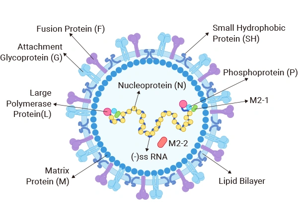
Figure 1. Schematic structural diagram of respiratory syncytial virus
RSV’s genome is a linear single-stranded negative-sense RNA strand approximately 15.2 kb long, containing 10 independent open reading frames (ORFs). These coding regions produce 11 functional proteins, split into structural and non-structural categories, each undertaking distinct roles in viral replication, particle assembly, and infectious progression. The detailed functions of each protein are listed below:
Protein Functional Table
| Protein Names | Core Biological Function |
|---|---|
| Fusion Protein (F) | Mediates membrane fusion between virions and host cell membranes to facilitate viral entry. It serves as a key antigenic epitope that induces neutralizing antibody production, representing the primary target for protective immune responses and antiviral therapeutic development. |
| Attachment Protein (G) | Mediates initial viral binding to host cell surfaces to initiate infection. The G protein exhibits high sequence variability, which defines antigenic epitope diversity and acts as a major biomarker to distinguish RSV subtype A and subtype B strains. |
| Small Hydrophobic Protein (SH) | A short transmembrane protein with partial involvement in viral infection and replication cascades, though its functional significance is less prominent compared to F and G glycoproteins. |
| Nucleoprotein (N) | Associates tightly with viral genomic RNA to form intact nucleocapsids, shielding viral genetic material from degradation. |
| Phosphoprotein (P) | Forms the viral replication complex alongside N protein and large L polymerase, participating in all stages of viral genome replication. |
| RNA Polymerase Protein (L) | Carries intrinsic RNA-dependent RNA polymerase catalytic activity, responsible for viral RNA transcription and full-length genome replication. |
| Matrix Protein M1 (M) | Situated directly beneath the lipid envelope, links membrane glycoproteins to internal RNP complexes and coordinates viral particle assembly. |
| Matrix Protein M2-1 (M2-1) | Cooperates with M protein during virion assembly and budding; additionally modulates the efficiency of viral RNA synthesis. |
| Matrix Protein M2-2 (M2-2) | Regulates viral transcription and replication cycles, tuning overall viral replication efficiency by adjusting RNA production levels. |
| Non-structural Protein NS1 | Regulatory factor modulating viral replication and infectivity; its complete functional mechanisms remain partially uncharacterized. |
| Non-structural Protein NS2 | Also participates in modulating viral replication and infection pathways, with its precise molecular mechanisms still under ongoing research. |
RSV infection proceeds through five sequential stages: surface attachment, cellular entry, intracellular genome replication, virion assembly, and extracellular release:
2.1 Viral Attachment
RSV spreads via respiratory aerosol droplets and first targets epithelial cells lining the human upper respiratory tract. Viral surface G glycoproteins recognize and bind specific receptor molecules on host cell membranes, predominantly heparan sulfate proteoglycans (HSPGs) plus several auxiliary receptor candidates.
2.2 Viral Entry
After stable attachment, conformational rearrangement of the F glycoprotein triggers fusion between the viral envelope and host cell plasma membrane, allowing internalization of the viral nucleocapsid. RSV can also enter host cells via clathrin-mediated endocytosis: virions are wrapped into cytoplasmic endosomes, where acidic luminal conditions activate the F protein to drive fusion between viral and endosomal membranes, releasing the RNP core into the host cytoplasm.
2.3 Viral Replication
Once inside the cytoplasm, the viral nucleocapsid disassembles to expose the negative-sense RNA genome. Viral RNA-dependent RNA polymerase (composed of L and P subunits) reads the genomic template and generates multiple positive-sense messenger RNAs. These mRNAs are translated by host ribosomes to produce all viral structural and non-structural proteins. The polymerase complex also generates full-length positive-sense RNA intermediates, which act as templates for synthesizing new progeny negative-sense viral genomes to be packaged into nascent virions.
2.4 Viral Assembly and Release
Newly replicated viral genomic RNA binds to N protein to assemble fresh nucleocapsid cores. These RNPs migrate toward the host cell plasma membrane, where they interact with cytoplasmic domains of the M matrix protein. M protein recruits membrane-anchored G and F glycoproteins to construct complete viral particles. Mature virions bud outward through the host cell membrane, acquiring a segment of host lipid bilayer to form their outer envelope before release into the extracellular space.
2.5 Host Immune Response & Tissue Pathology
RSV infection simultaneously activates innate and adaptive immune defense pathways in the human body. The innate immune response drives interferon secretion and abundant pro-inflammatory mediator release, while adaptive immunity generates RSV-specific neutralizing antibodies and antigen-specific T cell populations. Viral replication destroys airway epithelial cells, triggers heavy inflammatory cell infiltration, increases airway mucus secretion, narrows respiratory tracts, and elevates bronchial hyperreactivity. These tissue injuries induce clinical manifestations including persistent cough, wheezing, dyspnea, and in severe cases, progress to acute bronchitis or viral pneumonia.
Comprehensive understanding of RSV’s full infectious cycle provides a critical theoretical foundation for developing effective preventive vaccines and targeted antiviral therapeutics. Current research programs for RSV vaccines and anti-viral agents continue to advance, aiming to curb RSV transmission and mitigate its pathogenic damage by interfering with key steps in the viral life cycle.
Global research into RSV antiviral compounds mainly focuses on four core therapeutic targets: the fusion F protein, viral RNA-dependent RNA polymerase (RdRp), host cell dependency factors, and the deoxyguanosine nucleotide biosynthetic pathway. Research surrounding these targets lays essential groundwork for developing novel anti-RSV small-molecule drugs.
3.1 Fusion Protein (F Protein)
The F glycoprotein is an indispensable mediator enabling RSV penetration into host cells by facilitating membrane fusion, making it a leading target for antiviral drug development. Compounds such as GS-5806 and BMS-433771 block F protein conformational changes to inhibit virion-host cell fusion and prevent infection. Furthermore, RV521, a small-molecule RSV-F inhibitor, has demonstrated potent therapeutic effects in human clinical trials, lowering viral load and alleviating clinical disease severity.
3.2 RNA-Dependent RNA Polymerase (RdRp)
RSV’s viral polymerase represents another high-value therapeutic target. Blocking its catalytic activity suppresses viral genome replication and halts the entire viral life cycle. Using structural bioinformatics design, scientists have identified novel allosteric binding pockets on the RSV RdRp complex, enabling the creation of potent small-molecule polymerase inhibitors with broad anti-RSV activity.
3.3 Host Dependency Factors
In recent years, an increasing volume of research prioritizes host-targeted antiviral strategies. Viral structural proteins N and G are validated functional targets due to their central roles during infection. Nucleolin has also been identified as an essential host receptor for RSV; small molecules binding its RNA-binding domain effectively restrict RSV cellular entry and replication.
3.4 Deoxyguanosine Nucleotide Synthesis Pathway
Multiple studies confirm that disrupting the host cell’s deoxyguanosine nucleotide synthesis cascade can robustly suppress RSV replication. Probenecid, an FDA-approved clinical medication, inhibits RSV propagation by blocking organic anion transporter (OAT) activity on host cell membranes.
| Target Protein | Full Product Name | Expression System | Tag Label Information |
|---|---|---|---|
| N | Recombinant Bovine respiratory syncytial virus Nucleoprotein (N) | E.coli | N-terminal 6xHis-SUMO-tagged |
| F | Recombinant Human respiratory syncytial virus A Fusion glycoprotein F0 (F), partial fragment | E.coli | N-terminal 6xHis-B2M-tagged |
| F | Recombinant Human respiratory syncytial virus A Fusion glycoprotein F0 (F), partial fragment | E.coli | N-terminal 6xHis-tagged |
| F | Recombinant Human respiratory syncytial virus A Fusion glycoprotein F0 (F), partial fragment | Yeast | N-terminal 6xHis-tagged |
| G | Recombinant Human respiratory syncytial virus A Major surface glycoprotein G (G), partial fragment | Baculovirus | N-terminal 10xHis-tagged + C-terminal Myc-tagged |
| G | Recombinant Human respiratory syncytial virus A Major surface glycoprotein G (G), partial fragment | E.coli | N-terminal 6xHis-SUMO-tagged |
| G | Recombinant Human respiratory syncytial virus A Major surface glycoprotein G (G), partial fragment, Biotinylated | E.coli | N-terminal 6xHis-SUMO3-Avi-tagged |
| F | Recombinant Human respiratory syncytial virus A Fusion glycoprotein F0 (F), partial fragment | E.coli | N-terminal 10xHis-tagged + C-terminal Myc-tagged |
| G | Recombinant Human respiratory syncytial virus B Major surface glycoprotein G (G), partial fragment | E.coli | N-terminal 6xHis-tagged |
| N | Recombinant Human respiratory syncytial virus B Nucleoprotein (N) | E.coli | N-terminal 10xHis-tagged + C-terminal Myc-tagged |
| F | Recombinant Human respiratory syncytial virus B Fusion glycoprotein F0 (F), partial fragment | Yeast | N-terminal 6xHis-tagged |
| M2-1 | Recombinant Human respiratory syncytial virus B Matrix M2-1 (M2-1) | E.coli | N-terminal 6xHis-tagged + C-terminal 6xHis-tagged |