
The Epstein-Barr virus (EBV), alternatively designated human herpesvirus 4, falls under the herpesvirus family and ranks among the most prevalent viruses that infect humans. Distributed globally, this pathogen frequently triggers viral pharyngitis, with young adults representing the highest-risk population. A vast majority of individuals will contract EBV at some stage of their lifespan, often without developing any noticeable clinical manifestations. Its primary transmission medium is bodily secretions, especially saliva. EBV is capable of inducing infectious mononucleosis (commonly shortened to mono) alongside an array of other clinical disorders.
This article covers comprehensive EBV-related knowledge, including viral structural features, key viral proteins, infection mechanisms, clinical manifestations, transmission pathways, laboratory diagnostic approaches and clinical management strategies.
EBV virions measure between 122 and 180 nanometers in diameter, housing a double-stranded DNA helix with roughly 172,000 base pairs encoding 85 distinct viral genes. The genomic DNA is wrapped within a protein nucleocapsid layer, which is further enclosed by a protein-rich tegument compartment. The outermost layer consists of a lipid-containing viral envelope studded with glycoprotein surface spikes; these glycoproteins are essential for the virus to attach and penetrate target host cells.
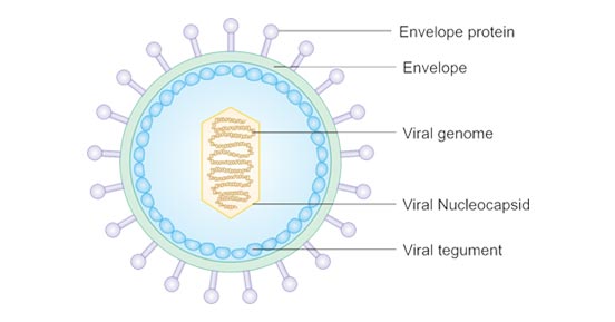
Figure 1: Schematic diagram illustrating the layered structure of EBV particles
| Target Gene Locus | Uniprot Accession ID | Protein Full Name | Functional Explanation |
|---|---|---|---|
| BALF5 | P03198 | DNA polymerase catalytic subunit | Responsible for replicating viral genomic DNA during the late lytic infection phase, generating long tandem concatemeric DNA fragments. |
| BCRF1 | P03180 | Viral interleukin-10 analog | Facilitates immune evasion of infected cells by blocking recognition mediated by cytotoxic T lymphocytes. |
| BDLF3 | P03224 | Glycoprotein BDLF3 | Viral envelope glycoprotein component |
| BHRF1 | P03182 | Apoptosis regulatory protein BHRF1 | Classified as early antigen R, one of EBV’s early expressed antigens |
| BLLF1 | P03200 | Envelope glycoprotein GP350 | Also referred to as EBV membrane antigen. It binds to the CR2 receptor expressed on B lymphocyte surfaces and promotes the interaction between viral gp42 glycoprotein and host HLA class II molecules. |
| BLLF3 | K9US42 | Deoxyuridine 5'-triphosphate nucleotidohydrolase | Participates in cellular nucleotide metabolism, preventing uracil residues from being incorporated into replicating viral DNA. |
| BRLF1 | Q3KSS7 | Replication and transcription activator protein | An immediate-early transcription factor governing the initiation of lytic viral gene expression and mediating lytic reactivation from latent EBV reservoirs. |
| BZLF1 | P03206 | Trans-activator protein BZLF1 | Functions as a dual-functional transcription factor: it activates early lytic cycle genes and acts as an origin-binding protein to support viral genome replication. |
| EBNA1 | P03211 | Epstein–Barr nuclear antigen 1 | EBNA-1 attaches to the oriP replication origin within the viral genome, controlling episome replication and segregation during host cell mitosis. It is the sole viral protein expressed during type I latency. |
| EBNA2 | Q69022 | Epstein-Barr virus nuclear antigen 2 | Serves as EBV’s primary transactivator; it modulates latent viral transcription and drives the permanent immortalization of EBV-infected host cells. |
| EBNA3 | P12977 | Epstein-Barr virus nuclear antigen 3 | A standard EBV genome encodes three homologous proteins of this family: EBNA-3A (EBNA-3), EBNA-3B (EBNA-4), EBNA-3C (EBNA-6). All three proteins can bind to the host cellular RBP-Jκ protein. |
| EBNA4 | P03203 | Epstein-Barr nuclear antigen 4 | Homologous latent nuclear antigen family member |
| EBNA6 | P03204 | Epstein-Barr nuclear antigen 6 | Homologous latent nuclear antigen family member |
| gB | P03188 | Envelope glycoprotein B | Forms spike protrusions on the outer virion envelope; mediates membrane fusion between viral and host cell membranes to facilitate viral entry. |
| gH | P03231 | Envelope glycoprotein H | The gH-gL heterodimer complex is mandatory for the fusion reaction between viral envelopes and host plasma membranes to complete cell entry. After binding host integrin molecules, gL dissociates from gH, triggering activation of fusion effector glycoproteins gB and gH. |
| gL | P03212 | Envelope glycoprotein L | Forms functional heterodimer with glycoprotein H |
| LMP1 | P13198 | Latent membrane protein 1 | A six-transmembrane spanning protein that is indispensable for EBV-induced cell proliferative transformation. |
| LMP2 | P13285 | Latent membrane protein 2 | LMP-2A and LMP-2B isoforms are transmembrane proteins that suppress intracellular tyrosine kinase signaling cascades. |
| SCP | P14348 | Small capsomere-interacting protein | Coats the outer surface of the capsid shell to assist intact infectious virion assembly, creating an intermediate layer separating the nucleocapsid and tegument. |
EBV employs distinct infection pathways to invade two primary target cell populations within the human body: B lymphocytes and epithelial cells.
For B lymphocyte infection: Viral surface glycoprotein gp350 first attaches to cell-surface receptors. Next, viral gp42 interacts with host MHC class II molecules, triggering fusion between the viral lipid envelope and the target cell membrane to enable EBV internalization. Human CD35 acts as an auxiliary attachment cofactor for gp350/220, creating an alternative infection route for CD21-negative cell subsets such as immature B cells. Once the acute primary lytic infection is suppressed by the host immune system, EBV establishes lifelong latent residence within the individual’s B cell pool.
For epithelial cell infection: Viral protein BMRF2 engages cellular β1 integrins as the initial binding step. Subsequently, the viral gH/gL complex binds to epithelial αvβ6 or αvβ8 integrin receptors. This molecular interaction initiates envelope-cell membrane fusion, allowing EBV virions to enter epithelial cells.
Saliva is the dominant transmission vector for EBV. Viral particles contained in saliva spread through intimate kissing, shared beverages and meals, communal tableware, toothbrushes, or contact with children’s saliva-contaminated toys. The virus can remain viable on contaminated surfaces as long as the material maintains moisture.
Non-salivary transmission pathways also exist: EBV may spread via blood and seminal fluid during sexual activity, allogeneic blood transfusions, and solid organ transplantation procedures.
Serological blood testing detecting EBV-specific antibodies serves as the standard diagnostic method, targeting three core antigen groups:
1. Viral capsid antigen (VCA): Anti-VCA IgM antibodies emerge rapidly during acute infection and disappear within several weeks, while anti-VCA IgG persists for the patient’s entire lifetime.
2. Early antigen (EA): Anti-EA antibodies develop during active viral replication; they generally become undetectable within months, though a subset of patients retain detectable levels long-term.
3. EBV nuclear antigen (EBNA): Anti-EBNA antibodies accumulate gradually over months post-infection and remain detectable throughout the patient’s lifespan.